Corneal Sensitivity
Corneal Nerves:
The Foundation of Ocular Surface Health
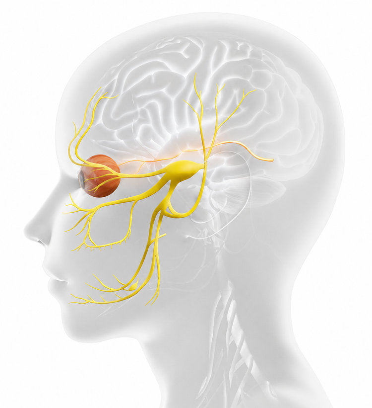
The cornea is the human tissue with the highest density of free nerve fiber endings.
The corneal nerves are essential for the ocular surface health, as they trigger 3 key ocular protection mechanisms:

Stimulation of corneal nociceptors by various factors activates neural pathways via the trigeminal nerve (cranial nerve V), relaying signals through the thalamus or cortex. This triggers the lacrimal and meibomian glands, along with conjunctival goblet cells, to secrete aqueous, lipid, and mucin components essential for natural tear production. Corneal nerve injury can follow many threats including mechanical/chemical trauma, infections, and surgeries. Corneal nerves main function is to protect the ocular surface and preserve corneal transparency and vision.
Corneal Sensitivity Impairment in Ocular Conditions
Corneal nerve health can be evaluated by analyzing the function (by esthesiometry or corneal sensitivity testing) or the anatomy and morphology (by in vivo confocal microscopy, IVCF).
Corneal sensitivity impairment (hypersensitivity or hyposensitivity) can compromise the ocular surface healing process and decrease tear production, increasing the risk of corneal deterioration and vision impairment. Corneal sensitivity can be evaluated using an esthesiometer, which allows to quantitatively measure corneal nerve function, allowing timely detection of potential ocular surface damage that requires prompt treatment.
A significant reduction in corneal sensitivity has been reported in multiple ocular conditions:
- Neurotrophic Keratitis
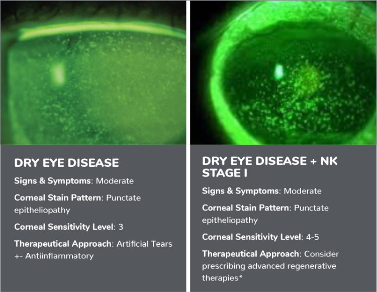
- Dry Eye Disease
- Diabetes
- Anterior Segment Surgery
- Contact Lens
- Glaucoma
Non-Contact Corneal Sensitivity Testing
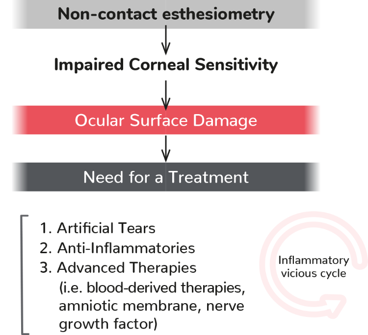
Non-contact esthesiometry is the test for screening, and monitoring of patients at risk for neurotrophic dysfunction associated with ocular surface disease.
When corneal sensitivity is impaired, neurotrophic damage occurs on the ocular surface that requires advanced therapies.
If Altered corneal sensitivity involves corneal nerve dysfunction that can lead to the development of epithelial defects, ulcerations, corneal perforations and reduced corneolimbic stem cell function.
Corneal Sensitivity Testing Clinical Applications
Non-contact esthesiometry is the test for screening, and monitoring of patients at risk for neurotrophic dysfunction associated with ocular surface disease.
When corneal sensitivity is impaired, neurotrophic damage occurs on the ocular surface that requires advanced therapies.
If Altered corneal sensitivity involves corneal nerve dysfunction that can lead to the development of epithelial defects, ulcerations, corneal perforations and reduced corneolimbic stem cell function.
1. Lum E, Corbett M, Murphy P. Corneal Sensitivity After Ocular Surgery. Eye & Contact Lens. 2019;00:1–12. 2. Yang AY, et al. Corneal Innervation and Sensation: The Eye and Beyond. Yale J Biol Med. 2018 Mar 28;91(1):13-21. eCollection 2018 Mar. 3. Patel S, et al. How Should Corneal Nerves be Incorporated Into the Diagnosis and Management of Dry Eye, Curr Ophthalmol Rep. 2021 Sep; 9(3): 65-76. 4. Tsubota K, et al. Defining Dry Eye from a Clinical Perspective. Int. J. Mol. Sci. 2020, 21. Vereertbrugghen A, et al. Corneal nerves and their role in dry eye pathophysiology, Experimental Eye Research, 2022; Volume 222, 109191. 5. Belmonte C, et al. TFOS DEWS Il pain and sensation report. Ocul. Surf. 2017;15:404-437. 6. Craig JP, et al. TFOS DEWS III Diagnostic Methodology report. Ocul Surf. 2024;32:1-101. doi:10.1016/j.jtos.2024.08.001. 7. Al Aqaba MA, Dhillon VK, Mohammed I, et al. Corneal nerves in health and disease, Prog Retin Eye Res. 2019;73:100762. 8. Jaenen N, et al. Ocular symptoms and signs with preserved and preservative-free glaucoma medications. Eur J Ophthalmol. 2007;17(3):341-9. 9. Villalba, M, . et al. Detection of Subclinical Neurotrophic Keratopathy by Noncontact Esthesiometry. Ophthalmol. 10. Ther 13, 2393–2404 (2024). https://doi.org/10.1007/s40123-024-00998-9. 11. Shao CG, et al. Novel Therapies for the Prevention of Fibrosis in Glaucoma Filtration Surgery. Biomedicines. 2023 Feb 21;11(3):657. doi: 10.3390/biomedicines11030657. 12. Surico PL, et al. Effects of Diabetes Mellitus on Corneal Immune Cell Activation and the Development of Keratopathy. Cells. 2024; 13(6):532. 13. Kolkedi Z, et al. Pre-Oph-thalmoscopic Quantitative Biomarkers in Diabetes Mellitus. Transl. Vis. Sci.Technol. 2023;12(3). Ljubimov AV. Diabetic complications in the cornea. Vision Res. 2017;139:138-152. 14. Rosenberg ME, et al. Corneal structure and sensitivity in type 1 diabetes mellitus. Invest Ophthalmol Vis Sci. 2000;41(10):2915-2921. 15. Edwards K, et al. Standardized corneal esthesiometry in diabetic patients. Cornea. 2012;31(4):401-407. 16. Mvilongo C, et al. Clinical profile of corneal sensitivity in diabetic patients: A case-control study. Journal Francais D’ophtalmologie.2024 Sep;47(7):104212. DOI: 10.1016/j.jfo.2024.104212. PMID: 38788250. 17. Bandeira F, et al. Corneal re-innervation following refractive surgery treatments. Neural Regen Res. 2019 Apr; 14(4): 557-565. 18. Dohlman TH, et al. Lai EC, Ciralsky B. Dry eye disease after refractive surgery. Int Ophthalmol Clin. 2016;56: 101-110. 19. Algorithm presented at the Annual Congress of the Andalusian Society of Ophthalmology and at the Annual Congress of Facoelche 2024 by Dr. Salvador Garcia Delpech. 20. Stapleton F, et al. Topical Review: Effects of Contact Lens Wear on Corneal, Conjunctival, and Lid Margin Sensitivity. Optom Vis Sci. 2019;96(10):790-801. doi:10.1097/OPX.0000000000001429. 21. Stapleton F, et al. The TFOS International Workshop on Contact Lens Discomfort: report of the subcommittee on neurobiology. Invest Ophthalmol Vis Sci. 2013;54(11):TFOS71-TFOS97. Published 2013 Oct 18. doi:10.1167/iovs.13-13226. 22. Mastropasqua L, Massaro-Giordano G, Nubile M, Sacchetti M. Understanding the pathogenesis of neurotrophic keratitis: the role of corneal nerves. J Cell Physiol. 2017;232(4):717-724. 23. Adapted Contact Esthesiometry. Res Sq [Preprint]. 2023 May 22:rs.3.rs-2833826. doi:10.21203/rs.3.rs-2833826/v1. PMID: 37292809.