
The first non-contact portable medical device to assess ocular surface health by monitoring corneal nerve function
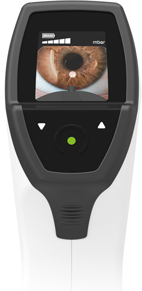
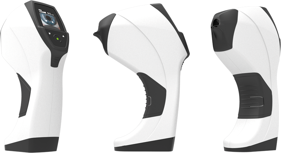
NEW TECHNOLOGY FOR THE MOST COMPLETE EYE EXAM
For screening patients at risk of Neurotrophic Keratitis, allowing early intervention:
· Dry Eye Disease
· Contact lens
· Diabetes
· Refractive and Cataract surgery
· Glaucoma
Key features
Contactless
Non-invasive and safe technology
Portable
Quick and easy to use, designed for appropriate placement in the slit lamp.
Innovative airflow technology
Uses a patented flow technology and a high-precision electronic LED positioning system.
Five standardized stimulus levels
They help to classify corneal sensitivity into different categories, serving as a guide to the degree of impact on corneal nerve function.
Replicable results outside the operator
For consistent and reliable patient follow-up.
No consumables required
Contactless
Non-invasive and safe technology
Portable
Quick and easy to use, designed for appropriate placement in the slit lamp.
Innovative airflow technology
Uses a patented flow technology and a high-precision electronic LED positioning system.
Five standardized stimulus levels
They help to classify corneal sensitivity into different categories, serving as a guide to the degree of impact on corneal nerve function.
Replicable results outside the operator
For consistent and reliable patient follow-up.
No consumables required
Non-contact esthesiometry
| Stimulus Level - Threshold | Corneal sensitivity1,2 |
|---|---|
|
Level 1 |
Suspected hypersensitivity |
|
Level 2 |
Normal corneal sensitivity |
|
Level 3 |
|
|
Level 4 |
Mild corneal hyposensitivity |
|
Level 5 |
Severe corneal hyposensitivity |
|
No response at any level (Level 6) |
Very severe corneal hyposensitivity |
1. Merayo-Lloves J, Gómez Martín C, Lozano-Sanroma J, Renedo Laguna C. Assessment and safety of the new esthesiometer BRILL: Comparison with the Cochet-Bonnet Esthesiometer. Eur J Ophthalmol. 2024 Jul;34(4):1036-1045. 2. Ruiz-Lozano RE, Quiroga-Garza ME, Ramos-Dávila EM, Pantaleón-García J, Khodor A, Komai S, et al. Comparative Evaluation of the Corneal Sensitivity Thresholds between the Novel Non-Contact and Cochet-Bonnet Esthesiometers. Am J Ophthalmol. 2025 Mar;271:407-416.
Worldwide presence of corneal esthesiometer Brill
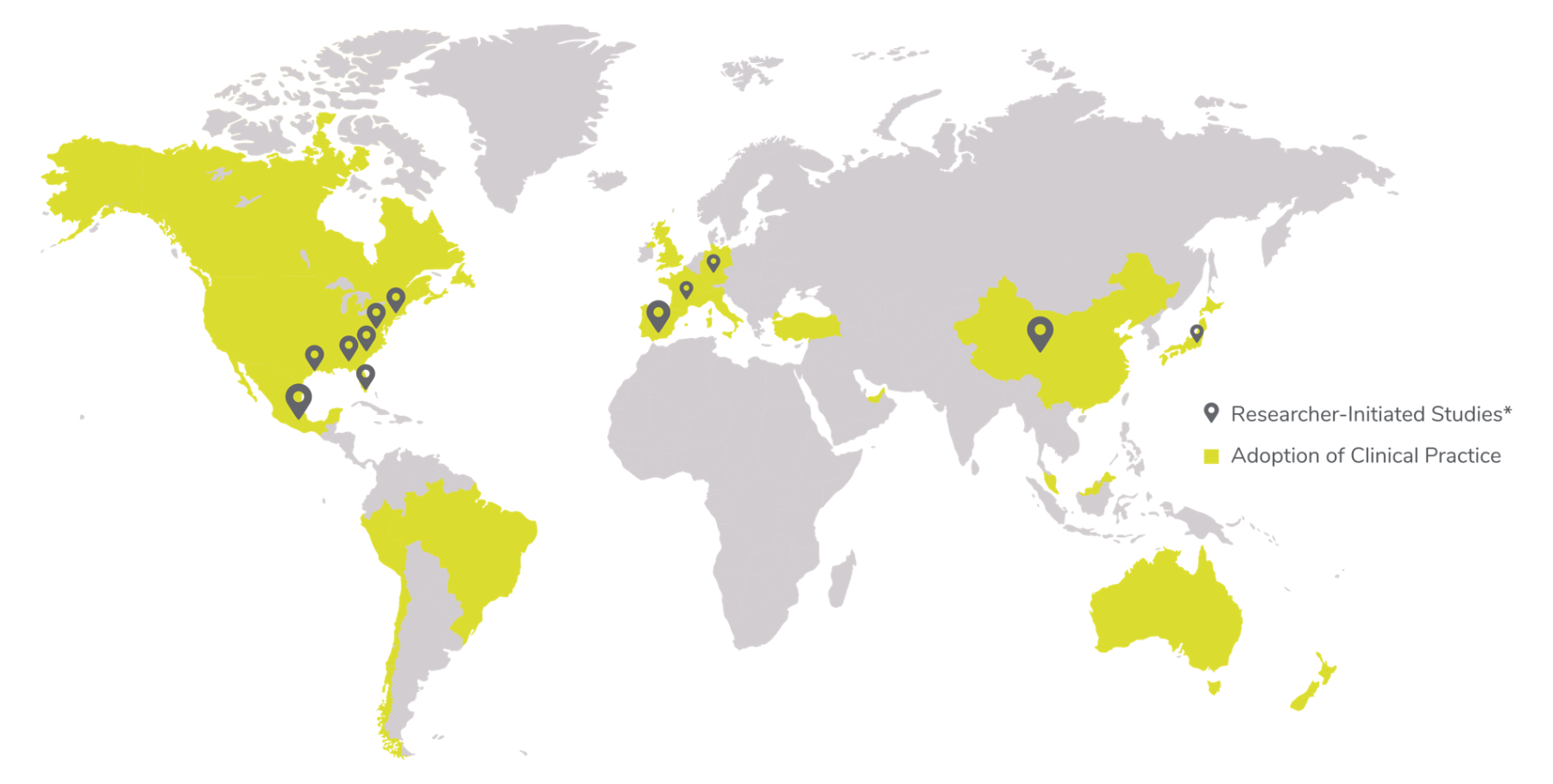
*Ongoing clinical research initiated by healthcare professionals to date, not sponsored by Brill.
Non-contact esthesiometry
Effective and accurate diagnosis of neurotrophic keratitis and corneal neurotrophic dysfunction in ocular conditions
Early detection of NK stage I. Monitoring of Corneal Neurotrophic disfunction in patients at risk, with ocular conditions such as DED, Diabetes, Glaucoma, Contact lens use, refractive and cataract surgery, etc. Detection of different functional states of the cornea using 5 validated levels of Corneal Sensitivity Testing.
Patient monitoring for optimum therapeutic adjustements
Useful in assessing the efficacy of therapies for dry eye disease, corneal nerve regeneration, ocular surface diseases, and diabetic neuropathy. Provides valuable corneal sensitivity metrics to complement other ocular assessments.
Patient selection for dry eye disease & nk clinical trials
Accurate screening of DED and/or NK patients following established inclusion and exclusion criteria for clinical trials.
Assessment of study drug cornea-related adverse events
Evaluation of systemic or local study drug side effects related to general ocular surface ophthalmic damage and/or specific corneal toxicity effects.
Non-contact esthesiometry
Effective and accurate diagnosis of neurotrophic keratitis and corneal neurotrophic dysfunction in ocular conditions
Early detection of NK stage I. Monitoring of Corneal Neurotrophic disfunction in patients at risk, with ocular conditions such as DED, Diabetes, Glaucoma, Contact lens use, refractive and cataract surgery, etc. Detection of different functional states of the cornea using 5 validated levels of Corneal Sensitivity Testing.
Patient monitoring for optimum therapeutic adjustements
Useful in assessing the efficacy of therapies for dry eye disease, corneal nerve regeneration, ocular surface diseases, and diabetic neuropathy. Provides valuable corneal sensitivity metrics to complement other ocular assessments.
Patient selection for dry eye disease & nk clinical trials
Accurate screening of DED and/or NK patients following established inclusion and exclusion criteria for clinical trials.
Assessment of study drug cornea-related adverse events
Evaluation of systemic or local study drug side effects related to general ocular surface ophthalmic damage and/or specific corneal toxicity effects.
Advantages of integrating the brill corneal esthesiometer into routine eye examinations
Early detection of corneal nerve dysfunction (both hypo- and hypersensitivity), allowing intervention before irreversible ocular surface damage occurs.
- Improves diagnosis for neurotrophic keratopathy (NK) and other ocular surface diseases.
- Provides repeatable data to monitor disease progression and response to therapy over time.
- Guides treatment decisions, helping to optimize therapy for non-responder dry eye patients.
- Identifies candidates for advanced therapies (NGF, amniotic membrane, or autologous serum).
- Enhances patient outcomes and satisfaction through earlier and more targeted interventions.
- Elevates the clinic’s technological and diagnostic standards, reinforcing its position as a center of excellence in ocular surface care.
- Improves patient retention and trust, as sensitivity testing adds value and precision to follow-up visits.
- Creates opportunities for revenue increase, either through procedural billing or indirect value from increased procedures and patient visits.
Device applications for clinical trial applications
-
Accurate Diagnosis of Corneal Nerve Dysfunction
Enables screening and consistent early detection of neurotrophic keratitis in at-risk patients (DED, diabetes, glaucoma, AS surgery, CL fitting, Herpes Keratitis, Keratoconus, Sjögren…).
-
Patient Monitoring and Therapy Assessment
Repeatability and standardization support reliable patient monitoring for disease progression or regression, evaluation of treatment effects, and assessment of corneal nerve regeneration.
-
Clinical Trial Selection and Monitoring
-
Facilitates patient inclusion or exclusion for clinical trials (e.g. distinguishing between DED patients and those with a neurotrophic or neuropathic component)
-
Assist in monitoring efficacy of therapies (e.g. new treatments for nerve regeneration, neurostimulators, preservative-free therapies).
-
Assess potential drug-related adverse events on the cornea (e.g. oncology therapies that could cause corneal toxicity)
-
Data on file. BRILL PHARMA S.L. 2024.
1. Lum E, Corbett M, Murphy P. Corneal Sensitivity After Ocular Surgery. Eye & Contact Lens. 2019;00:1–12. 2. Yang AY, et al. Corneal Innervation and Sensation: The Eye and Beyond. Yale J Biol Med. 2018 Mar 28;91(1):13-21. eCollection 2018 Mar. 3. Patel S, et al. How Should Corneal Nerves be Incorporated Into the Diagnosis and Management of Dry Eye, Curr Ophthalmol Rep. 2021 Sep; 9(3): 65-76. 4. Tsubota K, et al. Defining Dry Eye from a Clinical Perspective. Int. J. Mol. Sci. 2020, 21. Vereertbrugghen A, et al. Corneal nerves and their role in dry eye pathophysiology, Experimental Eye Research, 2022; Volume 222, 109191. 5. Belmonte C, et al. TFOS DEWS Il pain and sensation report. Ocul. Surf. 2017;15:404-437. 6. Craig JP, et al. TFOS DEWS III Diagnostic Methodology report. Ocul Surf. 2024;32:1-101. doi:10.1016/j.jtos.2024.08.001. 7. Al Aqaba MA, Dhillon VK, Mohammed I, et al. Corneal nerves in health and disease, Prog Retin Eye Res. 2019;73:100762. 8. Jaenen N, et al. Ocular symptoms and signs with preserved and preservative-free glaucoma medications. Eur J Ophthalmol. 2007;17(3):341-9. 9. Villalba, M, . et al. Detection of Subclinical Neurotrophic Keratopathy by Noncontact Esthesiometry. Ophthalmol. 10. Ther 13, 2393–2404 (2024). https://doi.org/10.1007/s40123-024-00998-9. 11. Shao CG, et al. Novel Therapies for the Prevention of Fibrosis in Glaucoma Filtration Surgery. Biomedicines. 2023 Feb 21;11(3):657. doi: 10.3390/biomedicines11030657. 12. Surico PL, et al. Effects of Diabetes Mellitus on Corneal Immune Cell Activation and the Development of Keratopathy. Cells. 2024; 13(6):532. 13. Kolkedi Z, et al. Pre-Oph-thalmoscopic Quantitative Biomarkers in Diabetes Mellitus. Transl. Vis. Sci.Technol. 2023;12(3). Ljubimov AV. Diabetic complications in the cornea. Vision Res. 2017;139:138-152. 14. Rosenberg ME, et al. Corneal structure and sensitivity in type 1 diabetes mellitus. Invest Ophthalmol Vis Sci. 2000;41(10):2915-2921. 15. Edwards K, et al. Standardized corneal esthesiometry in diabetic patients. Cornea. 2012;31(4):401-407. 16. Mvilongo C, et al. Clinical profile of corneal sensitivity in diabetic patients: A case-control study. Journal Francais D’ophtalmologie.2024 Sep;47(7):104212. DOI: 10.1016/j.jfo.2024.104212. PMID: 38788250. 17. Bandeira F, et al. Corneal re-innervation following refractive surgery treatments. Neural Regen Res. 2019 Apr; 14(4): 557-565. 18. Dohlman TH, et al. Lai EC, Ciralsky B. Dry eye disease after refractive surgery. Int Ophthalmol Clin. 2016;56: 101-110. 19. Algorithm presented at the Annual Congress of the Andalusian Society of Ophthalmology and at the Annual Congress of Facoelche 2024 by Dr. Salvador Garcia Delpech. 20. Stapleton F, et al. Topical Review: Effects of Contact Lens Wear on Corneal, Conjunctival, and Lid Margin Sensitivity. Optom Vis Sci. 2019;96(10):790-801. doi:10.1097/OPX.0000000000001429. 21. Stapleton F, et al. The TFOS International Workshop on Contact Lens Discomfort: report of the subcommittee on neurobiology. Invest Ophthalmol Vis Sci. 2013;54(11):TFOS71-TFOS97. Published 2013 Oct 18. doi:10.1167/iovs.13-13226. 22. Mastropasqua L, Massaro-Giordano G, Nubile M, Sacchetti M. Understanding the pathogenesis of neurotrophic keratitis: the role of corneal nerves. J Cell Physiol. 2017;232(4):717-724. 23. Adapted Contact Esthesiometry. Res Sq [Preprint]. 2023 May 22:rs.3.rs-2833826. doi:10.21203/rs.3.rs-2833826/v1. PMID: 37292809.